Anatomical Pathology
The Anatomical Pathology Section at King Abdulaziz Medical City includes Surgical Pathology (Histopathology), and Cytology. There are currently eight highly trained, board certified and experienced Consultant Pathologists. Each is sub-specialized in at least one field of Anatomic Pathology. The subspecialties covered include GI and Liver Pathology, Lung Pathology, Neuropathology, Dermatopathology, Gynecological and Breast pathology and Renal Pathology. The section also has a number of rotating Anatomic Pathology Residents, Pre-scholar Residents and from time to time other clinical discipline Residents taking rotation in the Lab.
Histopathology:
Over 10,000 specimens are received yearly in the histopathology lab, and range from small biopsies to complex surgical specimens. The lab is operated by 10 qualified technical staff who deals with every facet of surgical specimen processing, and also actively involved in the training of students and laboratory members. There is a strong focus on the established quality systems and policies within the laboratory, to provide a high level of technical capabilities at all times.
This part of the lab offers many services, such as: Specimen Gross Photography, Rapid Frozen Section procedures providing provisional diagnosis within 20 minutes, technical support for renal biopsy procedures, and specialized collection requirements for Flow Cytometry, microbiological studies, molecular pathology and Immunofluorescence.
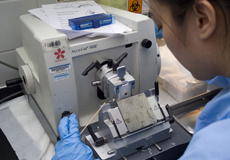
The Histology Laboratory has some of the finest equipment available, such as the 3 Sakura VIP Tissue Processor, the Leica CM1850, and CM1900 Cryostats for Tissue Frozen Section analysis, Modular Tissue-Tek Embedding Centers by Sakura, and Accu-cut Sakura Microtomes for sectioning.
Recent acquisitions have seen the Histology Laboratory excel in the area of Immunohistochemistry with the purchase of a state of the art automated staining module, the “Benchmark” manufactured by Ventana Medical Systems. Nearly, 100 antibodies are available for assisting difficult diagnosis and combined with this technology, quality and standardization is highly optimized.
Future plans for expansion of the laboratory are in progress, which will allow more specialized services to be available, such as Enzyme Histochemistry, In-situ hybridisation and molecular diagnostic techniques. Electron microscopy unit is coming soon to the lab, when the lab expansion is finalized.
Cytology:
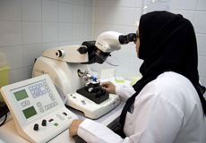
The Cytology Laboratory performs around 6000 Gynecology and Non-gynecology cases each year with close to 1000 fine-needle aspiration (FNA) biopsies. There are four Cytoscreeners and one Cytopreparatory technologists.
Many FNA’s are performed by Consultant Pathologists at the FNA Clinic and subsequent special ancillary studies, such as flow cytometric surface marker analysis, cytogenetic analysis and Immunocytochemistry, are available.
Recent New technology of liquid based cytology (SurePath) has been implemented for the Gynecology cases (Pap Smears). This technology provides more accurate results for Pap smears and offers the opportunity to perform various studies for sexually transmitted diseases, such as Human Papilloma Virus (HPV), Chlamydia and Neisseria gonorrhoeae.
Future visions for the Cytology Laboratory is to implement DNA testing of HPV and establish a cellular pathology training program for pathology residents, as well as a Cytotechnology school for technologists. In addition, the Cytology Laboratory is striving to be the national screening program for cervical cancer in the Kingdom.
Electron Microscopy Unit:
The Department of Pathology and Laboratory Medicine is pleased to announce that a new electron microscopy facility is now available. Electron microscopy allows the ultra structural study of various types of tissue, a process that proved to be essential in the diagnosis of large number of clinical conditions. It is expected that the newly established electron microscopy unit will be of a great value to evaluate tissue from renal biopsy, central and peripheral nervous systems, muscle biopsies, and various surgical specimens.
Additional special tests can also be performed using the electron microscopy such as examination of bone marrow aspirate, and cytological specimens. Physicians and scientists from all clinical specialties are encouraged to utilize the electron microscope. The average turn around time for an adequate specimen is 3-5 days.
The EM facility is an essential part of the Department of Pathology and Laboratory Medicine. Dr. Khaled Al Saad is in-charge to the unit while Mr. Saleem Baig (Supervisor- EM unit) and Ms. Hanouf Al Hamdan (EM Technologist) are responsible for electron microscopy operations.